Rare Primitive Neuroectodermal CancerousTumour Successfully Removed
Wednesday, February 25, 2009
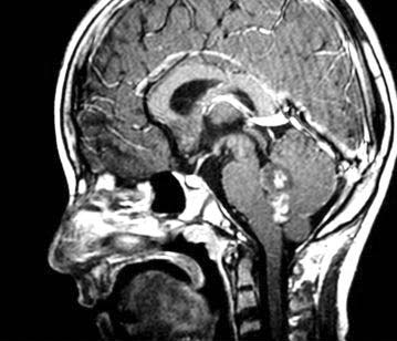
A team of 10 doctors at Wockhardt Hospitals Bangalore led by Dr. Shabeer Ahmed, Minimal Access Surgeon, Wockhardt Hospitals, successfully removed a cancerous tumor involving the abdominal aorta of a 19 year old girl, after a 7 hour long surgery. Saleema (name changed) travelled all the way from Jaipur to undergo the surgery at Wockhardt Hospitals,Bangalore to remove Primitive Neuroectodermal Tumour (PNET), a tumor that originates in cells from the Primitive Neural Crest. This cancerous tumor which involving abdominal aorta is a rare case in the medical history and there have been no reports found on such a rare tumor in the medical literature (reference – Cochrane Library –an international medical library, a collection of databases in medicine and other healthcare specialties provided by the Cochrane Collaboration and other organizations).
Most PNET’s occurs in the brain, extremities, pelvis, chest wall and mediastinum. This is the first quoted instance where a PNET was found in the abdomen involving the Bifurcation Abdominal Aorta. Surgical removal is usually a big challenge and it becomes more when it involves big vessels like the aorta – the big vessel which carries purified blood from heart to other organs.
Generally PNET is a rare tumor, usually occurring in children under 10 years old belongs to the Ewing family of tumors.The Ewing family of tumors is a group of cancers that includes Ewing tumor of bone (ETB or Ewing sarcoma of bone), extraosseous Ewing tumors (EOE tumors), primitive neuroectodermal tumors (PNET or peripheral neuroepithelioma), and Askin tumors (PNET of the chest wall). These tumors all come from the same type of stem cell.
During the surgery it was observed that the tumor had spread through the left-ureter, common aorta, bifurcation and the tumor was excised en-bloc along with the aorta.
According to Dr. Shabeer Ahmed, Minimal Access Surgeon, Wockhardt Hospitals, “Most PNET’s occurs in the brain, extremities, pelvis, chest wall and mediastinum. This is the first quoted instance where a PNET was found in the abdomen involving the Bifurcation Abdominal Aorta. Surgical removal is usually a big challenge and it becomes more when it involves big vessels like the aorta – the big vessel which carries purified blood from heart to other organs.”
Doctors give a 4 Hour Old New Heart and New Life

This was a first of its kind unique medical breakthrough which the Doctors at Wockhardt Hospitals found themselves to be a part of.The Pediatric Cardiac team at Wockhardt Hospitals, Mumbai at Mulund performed a life saving surgery on a 4 hour old baby gifting a life and in the process creating a new heart.
Pediatric Cardiac Consultants at Wockhardt Hospital have successfully performed a critical 2-hour beating heart surgery to allow the flow of blood from the baby’s heart to her lungs, thereby enabling the child to breathe normal. This life threatening, rare cardiac complication in neonates is called Pulmonary Atresia, that requires to be addressed in a critical window period of just a few hours of the baby’s birth, was accomplished in the state-of-the-art dedicated pediatric cardiac set up of the hospital.
A few minutes after her birth out of a planned caesarian section in a private city hospital the baby was shifted to Wockhardt Hospitals at Mulund .Within four hours of her birth, the baby was wheeled in for a beating heart BT Shunt procedure wherein a synthetic tube (shunt) was inserted to connect a new artery the sub clavian artery -- to the pulmonary artery. The sub clavian artery supplies blood to the arms but is now redirected to share supply to the arms and the lungs.
The child suffered from a rare congenital disorder called 'Pulmonary Atresia" where the the pulmonary valve of the heart which controls blood to the lungs was not developed and no blood was reaching the lungs. The child, however survived for a few critical hours due to ductus arteriosus, a valve that is meant for its fetal existence only. This valve shuts close naturally after birth and the corrective surgery was required within this window.
Pulmonary atresia is a congenital malformation of the pulmonary valve in which the valve orifice fails to develop. The valve is completely closed thereby obstructing the outflow of blood from the heart to the lungs.The only source of pulmonary blood flow is a patent ductus arteriosus. Due to this, the newborn baby is blue in color and pulmonary atresia can usually be diagnosed within hours or minutes after birth.
Foetal Echocardiography is used to diagnose heart defects before a child is born. The diagnosis done through foetal echocardiography helps in treating the heart defect in the child at an early stage especially when many times some interventions needs to be done immediately after the birth. Since foetal echocardiography was done in this case the defect was identified and immediately after her birth the baby was brought to Wockhardt Hospitals for treatment.”
In India, over 2.5 million children are born with heart defects every year and many lives are lost due to lack of proper infrastructure, awareness, and poor planning at time of birth. Some 3 per cent of these cases are of those born with Pulmonary Atresia. Timely detection and early treatment can not only save a child but also help him lead a normal and healthy life.
For more information and scheduling appointments with Doctors at Wockhardt Hospitals write into enquiries@wockhardthospitals.net
For more information and scheduling appointments with Doctors at Wockhardt Hospitals write into enquiries@wockhardthospitals.net
welcome to wockhardt Hospitals Blog
Welcome to the blog of Wockhardt Hospitals. We look forward to receiving your feedback and ideas on making this blog a truly representative of our online community
Subscribe to:
Posts (Atom)


