Doctor SpotLight :Cosmetic Surgeon ,Dr Apul Parikh
Sunday, May 17, 2009

Dr. Apul Parikh
Dip Cosmetic Surgery(London)
Ebopras(european boardof plastic reconstructive and aesthetic surgery)
MS, FRCS, Dip. Cosmetics
Wockhardt Hospitals, Bangalore.
Ebopras(european boardof plastic reconstructive and aesthetic surgery)
MS, FRCS, Dip. Cosmetics
Wockhardt Hospitals, Bangalore.
Today in Doctor Spotlight we feature Doctor Apul Parikh, who has recently joined us at Wockhardt Hospitals,Bannerghata Road as a Cosmetic Surgeon
Dr. Apul Parikh is amongst the leading plastic and cosmetic surgeons in India. Dr. Parikh has recently moved back to India from the United Kingdom. Dr. Apul initially graduated from the University Of Leeds School Of Medicine. After completing his basic surgical training he specialised in Plastic Surgery in which he has published a number of papers. He has worked in several prestigious London teaching hospitals, including the Royal Free Hospital, St. Bartholomew’s Hospital, the Royal London Hospital and Wexham Park Hospital. He also completed a fellowship at the Memorial Sloane-Kettering Cancer Hospital in New York. Dr Apul Parikh has recently passed the European Board of Plastic surgery (EBOPRAS).
Academic Training :Dr. Apul Parikh was actively involved in the teaching of plastic surgery to undergraduate and post-graduate trainees in the UK and was a Lecturer at the Royal Free Hospital and the Royal London Hospitals.
Dr. Apul Parikh has also received further super-specialised training in Cosmetic Surgery. He has successfully completed the UK’s maiden Diploma in Cosmetic Surgery and is now one of the lecturers to the students. He has been privileged to have been trained by Dr. Dai Davies, Dr. Nick Percival, Dr. Jan Stanek, and Dr. Simon Myers who are all regarded as the leading UK surgeons in the field of cosmetic surgery. Dr. Parikh is also one of the privileged few surgeons to have been trained in Cosmetic Vaginal Surgery.
More recently Dr. Parikh underwent specific training in Rhinoplasty (Nose jobs) in Dallas (USA) under the supervision of Dr. Rod Rohrich and Professor Jack Gunther. Dr. Apul Parikh is one of the few Cosmetic Surgeons to offer computer assisted consultations.
Cosmetic Surgery Procedures Offered by Dr Apul Parikh at Wockhardt Hospitals,Bangalore
Face:
▪ Hair transplantation, Brow lifts
▪ Facelift and Neck lift procedures
▪ Blepharoplasty (Upper and Lower Eyelid Surgery)
▪ Rhinoplasty (Nose Jobs)
▪ Otoplasty (Prominent ear correction)
▪ Lip Augmentation
▪ Injectible Fillers/Botox Injections
Breast Procedures
▪ Breast Augmentation
▪ Breast Reduction/Mastopexy (Uplift)
▪ Inverted Nipples Correction
▪ Gynaecomastia (Male Breast Reduction Surgery)
Body
▪ Abdominoplasty (Tummy Tucks), Thigh/Body Lift (Post Massive weight loss/Bariatric Surgery)
▪ Arm Reduction Surgery
▪ Liposuction
▪ Cosmetic Vaginal Surgery (Labia Reduction, Vaginal Tightening, G-Spot Augmentation)
Skin:
▪ Laser Surgery (Tattoo removal, Rejuvenation)
▪ Chemical Peels
▪ Spider Veins removal
▪ Scar Revision Surgery
For appointments with Dr Apul Parikh, please write to enquiries@wockhardthospitals.net
Patient Education Series:Treating Retinal Detachment
Friday, May 15, 2009
What is retina?
The retina is a thin, transparent tissue of light-sensitive nerve fibers & cells. It covers the interior wall of the eye like wallpaper covers the walls of a room. It functions like the film in a camera- light passes through the lens of the eye & is focused onto the retina. The retina “takes the picture” & transmits the image via the optic nerve to the brain.
What is retinal detachment?
It is a condition in which the light sensitive layer of the eye(retina) separates from the underlying eye wall & hence loses its function. It is a serious problem that may occur in any age although it usually occurs in middle-aged or older individuals. It is more likely to develop in people who are nearsighted (myopes) or in those whose relatives have had retinal detachments. A hard, solid blow to the eye may also cause this. If not treated early it may lead to impairment or loss of vision.
How does retinal detachment occur?
It is mainly caused by presence of one or more small tears or holes in the retina. Normal ageing can sometimes cause the retina to thin & degenerate (called lattice degeneration), but most often shrinkage of the vitreous body- the clear gel that fills the center of the eye, is responsible for the causation of retinal tears.
Shrinkage & detachment of the vitreous body is a common event with the age but does not cause any problem in most people. In few eyes that have abnormal strong attachments of the vitreous to the retina, a tear can result. Abnormal growth of the eye, which occurs in myopia or injury to the eye, may also cause the vitreous to shrink. Once a tear is present, watery fluid from the vitreous space may pass through the hole & flow between the retina & the outer wall of the eye. This seperates the retina & causes it to “detach” . The part of the retina that is detached does not function properly & there is a blur or a blind spot in vision.
There are other rare conditions such as tumor, which cause retinal detachment without formation of a hole/tear.
Retinal Detachment symptoms
One may see floating black spots called floaters, & flashes of light in the vision. In most cases, these do not indicate serious problems. But, in some cases, they may be associated with the retinal tears. A comprehensive eye examination by a retinal specialist to check the retina is necessary to determine if retinal tears are present. Such an examination is desirable as soon as symptoms develop because fresh retinal tears may be treatable without surgery, before they lead to a more severe retinal detachment.
Some retinal detachments may begin without noticeable floaters or flashes of light. In these cases, one may notice a wavy or watery quality in the overall vision or the appearance of a dark shadow in some part of the side vision. Further development of the retinal detachment will blur the central vision & create significant sight loss unless the detachment is repaired.
A few detachments may occur suddenly & one may experience a total loss of vision. Similar rapid loss of vision may also develop by bleeding in the vitreous, which happens when the retina is torn.
Diagnosing retinal detachmentA detachment retina cannot be viewed from the outside of the eye. Hence, if symptoms are noticed, a retinal surgeon should be visited as soon as possible. The specialist throughly examines the retina with an instrument called Indirect Opthalmoscope. The instruments bright light & magnification allows the specialist to locate areas of retinal tearing or weakness, which need to be corrected by treatment. Other special diagnostic instruments including special contact lenses, slit lamp etc & Ultrasound may also be used.
How to treat retinal detachmentThere is no medical treatment for retinal detachment. If the retina is torn & retinal detachment has not yet occurred, the same may be prevented by prompt prophylactic treatment. Once the retina becomes detached, it must be repaired surgically by a retinal surgeon. Successful re-attachment of the retina consists of sealing the retinal tear & preventing the retina from pulling away from the back of the wall of the eye again. There are several surgical procedures that may be used. The choice depends on the severity of the retinal detachment & the judgment of the surgeon.
There are 3 different techniques of treating Retinal Detachments1.Pneumatic retinopexy:
In very select group of patients, one may inject a gas bubble in the eye, treat the retinal hole with cryotherapy (freezing) or laser, & then position the eye to enable successful closer of the hole. This is the simplest of the treatment approaches with least intervention. The success of this procedure is about 70% & in case of failure, scleral buckling procedure can be done.
2.Scleral buckling: most other simple retinal detachments are handled by applying a silicone buckle on surface of the eye, thus indenting the walls inside. The retinal hole is treated with cryotherapy(freezing) & nthe fluid that has collected between the retina & the underlying layers is usually removed. The success rate of this surgery is usually 80-90%.
3.Vitreorentinal procedures: For more complex retinal detachments, complicated surgery called vitreoretinal surgery is needed. In this, the diseased vitreous is removed along with abnormal scar tissue. The retina is attached by use of air, gas or silicon oil. The success of these surgeries varies with type of case. Sometimes, multiple surgeries may be indicated in case of recurrence. Where silicone oil is used, it is usually removed after a variable period of time, once the retina is successfully reattached. The final success can be declared only if the retina remains attached after removal of silicone oil.
Obviously, the more complex the retinal detachment, the more complex will be the surgical procedure needed & less will be the cure rate.
Recovery and Post Operative Care:It is important to understand that surgical success & visual recovery need not go hand in hand. The visual recovery depends upon the basic strength in the retina, the duration of retinal detachment & most importantly, the health of the central, most sensitive part of the retina(mascula). Reading fine print needs excellent vision, hence only a percentage of the eyes with complex retinal detachment can regain reading capabilities. More often, mobile vision is retrieved. Failed surgery usually leads to non-recovery of vision & on occasions these eyes may shrink.
The surgery may be done under local or general anesthesia. With gas in the eye, air travel is restricted. Eye drops or ointment may have to be instilled for 6-8 weeks & glasses are prescribed at final examination.
With simple buckling surgery, vision may start recovering in a few days , but final vision is known after 6 weeks. With more complex vitreorentinal surgeries, it takes longer time for vision to improve & stabilize.
Patients with symptoms of retinal detachment require prompt attention by a retinal surgeon, who will throughly examine & advice about the need for treatment. It is important for persons with significant myopia or with family history of retinal detachment to have periodic eye examination. Early detection of changes in the vitreous or retina can be diagnosed & potential retinal detachment prevented.
To schedule an appointment with our eye care consultants at Wockhardt Hospitals please email us at enquiries@wockhardthospitals.net
To schedule an appointment with our eye care consultants at Wockhardt Hospitals please email us at enquiries@wockhardthospitals.net
Patient Education Series :Cataract Treatement
What is a Cataract?
A cataract is clouding of the normally clear lens of the eye. It can be compared to a glass window that is frosted or yellowed.
The amount and pattern of cloudiness within the lens can vary. If the cloudiness is not near the center of the lens, you may not be aware that a cataract is present.
The amount and pattern of cloudiness within the lens can vary. If the cloudiness is not near the center of the lens, you may not be aware that a cataract is present.
What are the Common symptoms of cataract
• A painless gradual blurring of vision
• Glare, or light sensitivity
• Poor night vision
• Double vision in one eye
• Needing brighter light to read
• Fading or yellowing of colors
What causes Cataract?
The most common type of cataract is age related to aging of the eye. Some other causes of cataract include:
• Family History
• Medical problems, such as diabetes
• Injury to the eye
• Medications, especially steroids
• Previous eye surgery
How fast does a cataract develop?
It is not possible to predict exactly how fast cataracts will develop in any given person. The development of cataract varies among individuals and may even be different between the two eyes.Most age-related cataracts progress gradually over a period of years.
Other cataracts, especially in younger people and people with diabetes, may progress rapidly over a short time.
His cataract treated?
Surgery is the only way a cataract can be removed. However, if symptoms of cataract are not bothering you very much, surgery may not be needed. Sometimes a simple change in your eyeglass prescription may be helpful.
No medications, dietary supplements or exercises have been shown to prevent or cure cataracts. Protection from excessive sunlight may help slow the progression of cataracts. Sunglasses that screen out ultraviolet (UV) light rays or regular eyeglasses with a clear, anti-UV coating offer this protection.
How is a cataract detected?
Your ophthalmologist can detect the presence of a cataract by performing a full eye examination.
A careful evaluation will also rule out any other conditions that may be causing blurred vision. Problems with other parts of the eye can be responsible for vision loss and may prevent you from having much or any improvement in vision after cataract surgery. If improvement in your vision is unlikely, cataract removal may not be recommended. Your eye doctor can tell you how much visual improvement is likely.
When should Cataract surgery be done?
Surgery should be considered when cataracts cause enough loss of vision to interfere with your daily activities.
It is not true that cataracts need to ripe before they can be removed or that they need to be removed just because they are present.
It is not true that cataracts need to ripe before they can be removed or that they need to be removed just because they are present.
Cataract surgery can be performed when your visual needs require it. You must decide if you can see well enough to do your job, drive safely and read and watch TV in comfort. Does your vision allow you to perform daily tasks, such as cooking, shopping, doing regular work or taking medications without difficulty?
Based on your symptoms, you and your ophthalmologist should decide together when surgery is appropriate.
What can I expect from cataract surgery?
More than 98% of those surgeries are performed successfully without complications.
During cataract surgery, which is usually performed under local or topical anesthesia as a daycare procedure, the cloudy lens is removed from the eye. In the most cases a permanent intra ocular lens implant(IOL) is placed in the eye.
Your ophthalmologist performs this delicate surgery using a microscope, miniature instruments and other modern technology. After surgery you will have to use eye drop as per your doctors advice.
Cataract surgery is a highly successful procedure. Improved vision is the result in over 98% of cases, unless there is a problem in other structures of the eye, It is important to understand that complications although rare can occur during or after the surgery.Cataracts are a common cause of decreased vision, particularly for the elderly, but they are treatable. Your ophthalmologist can tell you whether cataract or some other problem is the cause of your vision loss and can help you decide if cataract surgery is appropriate for you.
To schedule an online appointment with our Opthalmologist at Mumbai,please see below or and write to us at enquiries@wockhardthospitals.net
To schedule an online appointment with our Opthalmologist at Mumbai,please see below or and write to us at enquiries@wockhardthospitals.net
| Name of the Doctor | Outpatients Consultation Days | Timings |
| Dr.Ananad Subramanyam | Tue, Sat | 9 am to 12 pm |
| Dr.Hrihikesh Tadwalkar | Thur | 1pm to 2 pm |
| Dr.Nishikant Borse | Tue, Thu | 2 pm to 5 pm |
| Dr.C.M. Mavikar | Mon, Wed, Sat | 3 pm to 5 pm |
| Dr.Girija Suresh | Mon & Fri | 9 am to 1 pm |
| Tue, Thu, & Sat | 9 am to 5 pm | |
| Wed | 1 pm to 5 pm | |
| Dr.Jaganath Boramani | Tue | 3 pm to 5 pm |
| Dr.Nayan Thakkar | Thur | 5 pm to 7 pm |
| Dr.P.S.Suresh | Mon | 9 am to 7 pm |
| Tue Thur | 1 pm to 5 pm | |
| Wed | 9 am to 1 pm | |
| Fri | 1 pm to 7 pm | |
| Sat | 9 am to 5 pm | |
| Dr.Shivram Nadkarni | Tue & Sat | 10 am to 12 pm |
| Dr.Shashi Kapoor | Fri | 9 am to 1 pm |
| Wed | 7 pm to 9 pm | |
| Dr.Seeta Raman | Fri | 5 pm to 7 pm |
| Dr.Nitha Shanbag | Tue | 5 pm to 7 pm |
| Fri | 7 pm to 9 pm | |
| Dr.Swaranjit Singh Bhatti | Wed | 7 pm to 9 pm |
| Sat | 5 pm to 7 pm | |
| Dr.Manjiri Oak | Mon, Wed | 5 pm to 7 pm |
| Dr.Sumita Phatak | Mon, Wed, Fri | 9 am to 5 pm |
Study Shows Ginger capsules can lessen nausea if started days before cancer chemotherapy,
Thursday, May 14, 2009
Ginger capsules can lessen nausea if started days before cancer chemotherapy, study finds - CLTV: "Ginger capsules can lessen nausea if started days before cancer chemotherapy, study finds.
MARILYNN MARCHIONE AP Medical Writer reports
Ginger, long used as a folk remedy for soothing tummy aches, helped tame one of the most dreaded side effects of cancer treatment — nausea from chemotherapy, the first large study to test the herb for this has found.
People who started taking ginger capsules several days before a chemo infusion had fewer and less severe bouts of nausea afterward than others who were given dummy capsules, the federally funded study found.
'We were slightly beside ourselves' to see how much it helped, said study leader Julie Ryan of the University of Rochester in New York.
Results were released Thursday by the American Society of Clinical Oncology and will be presented at the group's annual meeting later this month.
Results were released Thursday by the American Society of Clinical Oncology and will be presented at the group's annual meeting later this month.
But don't reach for the ginger ale. Many sodas and cookies contain only flavoring — not real ginger, Ryan said. Her study tested a drug-like ginger root extract, and it's not known if people could get the same benefits from ginger teas or the powdered ginger sold as a spice.
Still, ginger capsules may offer a cheap, simple way to fight nausea, which is far more than just a quality-of-life issue, doctors say. Some cancer patients cut treatment short or refuse chemo altogether because of nausea, hurting their chances of beating the disease."
More Coverage here
More Coverage here
Nurses Day Celebrations at Bangalore
 Nurses at Wokhardt Hospitals,Bannerghata in a fashion show on International Nurses day
It was a Nurses day out on 12th of May at the Bannerghata Road Branch at Wockhardt Hospitals,Bangalore. For a changes the nurses at Wockhardt Hospitals donned the latest fashion couture and gave up their nursing uniform.
Not really couture or the essence of a fashion show,but the nurses showed why it is not that difficult sometimes to change to your roles and give something your best shot,if you have the passion.
Beginning with the lighting of the lamp, the International Nurses day celebrations,started after the inaugural address Dr Davison, Centre Head, Wockhardt Hospitals, Bannerghatta.
Mr. Davison congratulated all the nursing staff at Wockhardt on the occasion. “This is the noblest profession in the world probably the only profession that puts service before self,and not everyone can be a nurse. It requires a special calling, and the ability to be sympathetic, empathetic and truly concerned for the sick,” he said.
The show had three rounds and finally, the judges chose Dipty Diana Fernandes from Wockhardt, Cunningham Road, as “Queen of Wockhardt” and Rizwan from Wockhardt, Bannerghatta, as “King of Wockhardt”.
Wockhardt Hospitals is now on Twitter
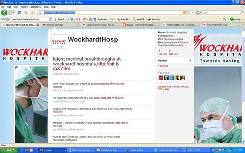
We welcome our users ,patients and caregivers to stay in touch with the latest news at wockhardt hospitals by following us on twitter. In the days to come we hope to use twitter to inform and update patients and caregivers about their friends and relatives ,who are admitted at Wockhardt hospitals and are eager to get an status update of their loved ones.
Our twitter url is http://twitter.com/wockhardthosp ! We welcome everyone to join us at twitter . Happy Twittering
Diabetes : The Number one silent Killer in India
Tuesday, May 12, 2009
According to the International Diabetes Federation the country with the largest numbers of people with diabetes is India (40.9 million), followed by China (39.8 million), the United States (19.2 million), Russia (9.6 million) and Germany (7.4 million).The World Health Organization projects that the number of diabetics will exceed 350 million by 2030.
In case you are not convinced about the enormity of the problem,Consider these statistics.
- One person is dying for every 10 seconds in the world.
- Two new diabetic cases are identifying for every 10 seconds in the world.
- 7 million new diabetic cases will be identified by 2025.
- 80% of diabetics in the world will be present in developing countries like India.
- India is the Diabetic capital of the world.
- It is not now a disease of rich people. It is a disease of sedentary people with Unhealthy diet habits.
In the USA
- 17.9m people are diagnosed with diabetes
- 5.7m people are undiagnosed with diabetes
- 57m people have pre-diabetes
- 186,300 (0.22%) people under 20 have diabetes
- 1 in every 400 to 600 under 20-year olds have Type 1 diabetes
- 2m adolescents have pre-diabetes
- 23.5m (10.7%) of those over 20 have diabetes
- 12.2m of those over 60 have diabetes
- 12m men (11.2%) have diabetes
- 11.5m women (10.2%) have diabetes
source:American Diabetes Association
Diabetes as it is sometimes believed, is not a disease of wealthy and rich alone. In fact almost two-thirds of the people with diabetes in our country are either of normal weight or even lean. Diabetes does not just happen overnight,and it does not segregate between the haves and have nots, it takes many years for diabetes to set in.That gives us a chance to early detection, possible prevention or even delaying it. Any person with, diabetes in family, obesity, physical or mental stress, needs to check blood glucose once a year after 30 years age.
At Wockhardt Hospitals, we recommended Diabetes screening at various stages of life, and for those with any of several risk factors. The screening test may be a random blood glucose test, a fasting blood glucose test, a blood glucose test a,two hours after 75 g of glucose, or an even more formal glucose tolerance test.The Details of the Preventive Diabetes Health Check Program are as follows
Comprehensive Diabetic health Check
HB,TC,DC,ESR
FBS
PPBS
Fasting Lipid profile
Serum Electrolytes
Serum calcium,P,Alkaline Phosphatase
Blood Urea,Serum, Creatinine/Potassium
HbA1c
Urine-Microalbuminuria ECG
Chest X Ray
Fundoscopy
Consultation with the Diabetologist/Dietician
Executive Diabetic Health Check
Fasting Lipid profile
Sr Electrolytes
HB,TC,DC,ESR
FBS
PPBS
Blood Urea
Sr Creatinine
Alkali Phosphatase
HbA1c
Urine Microalbuminuria
ECG
Chest X Ray
Fundoscopy
TMP
Echo
Consultation with Diabatelogist,Cardialogist & Dietician
Consultation with Physiotherapist
For details on our Diabetes Health Check Package please email us at enquiries@wockhardthospitals.net
International Nurses Day:Some People Deserve Much More than Just Thanks
Monday, May 11, 2009

Tomorrow is International Nurses day. 12th May is a special day for not only for those people who have led from the front in delivering care with a smile in their lips,but also a day for all of us in the medical fraternity who are so much dependent on them.
International Nurses Day is celebrated around the world every May 12,which is also the anniversary of Florence Nightingale's birth. The International Council of Nurses commemorates this important day each year along with a Nurses Day theme.
International Nurses Day is celebrated around the world every May 12,which is also the anniversary of Florence Nightingale's birth. The International Council of Nurses commemorates this important day each year along with a Nurses Day theme.
The International Nurses Day theme for 2009 is: Delivering Quality, Serving Communities: Nurses Leading Care Innovations.
The International Council of Nurses (ICN) has celebrated this day since 1965.
We at Wockhardt Hospitals would like to take this oppurtunity to thank our Nurses and would like to wish them a Happy Nurses day specialy for all those time we took your services for granted ..
Subscribe to:
Posts (Atom)


